Artificial intelligence is no longer a distant promise in dentistry. Cloud-based platforms capable of detecting caries, bone loss, and periapical lesions have received regulatory clearance from the US Food and Drug Administration — and are already being used in clinical practice. This post explores what these tools are, how they work, and what their arrival means for the future of dental diagnosis.
The diagnostic problem cloud AI is solving
Radiographic interpretation in dentistry is highly variable. Studies consistently show that interproximal caries, early bone loss, and periapical pathology are frequently missed or inconsistently graded across clinicians. This is not a skill problem — it reflects the inherent difficulty of reading two-dimensional projections of complex anatomy under time pressure.
A 2025 review in the International Dental Journal by Shujaat et al. — “FDA-Approved AI Solutions in Dental Imaging” — systematically evaluated all standalone, cloud-based AI platforms that have obtained FDA clearance for dental imaging. [1] The landscape is more mature than most clinicians expect.
How does it work?
The workflow is simple: the practice captures a radiograph as usual, uploads it to the platform, and receives an annotated report in under 10 seconds. All computation happens in the cloud — no local GPU required. The underlying models are convolutional neural networks (CNNs) trained on large annotated datasets of dental radiographs.
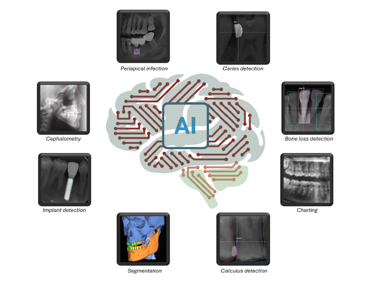
What can these tools detect?
| FINDING | IMAGE TYPE | EVIDENCE |
|---|---|---|
| Dental caries | Bitewing / periapical | Strong |
| Alveolar bone loss | Panoramic / periapical | Strong |
| Periapical lesions | Periapical | Strong |
| Calculus deposits | Bitewing | Moderate |
| Anatomical landmarks | CBCT / Panoramic | Emerging |
Performance and limitations
A 2024 meta-analysis found that AI models in dental imaging achieve an overall diagnostic accuracy of around 82%, comparable to — and sometimes exceeding — unassisted human examiners for specific tasks like interproximal caries detection.
That said, performance varies considerably by task and image quality. Dataset bias is a real concern — models trained on images from specific populations may underperform in different clinical settings. Explainability also remains limited: most tools return a bounding box or heatmap, but clinicians often need more context to trust and act on AI findings.
Why this matters
Coming from a clinical dental background and now studying health data science, what I find most compelling about this field is not the accuracy numbers — it is the accessibility argument. Cloud delivery means a small rural practice has access to the same analytical capability as a university hospital, with no hardware investment beyond a browser. The challenge now is not building better models, but building the evidence base and clinical trust needed for meaningful adoption.
References
[1] Shujaat S, Aljadaan H, Alrashid H, Aboalela AA, Riaz M. FDA-Approved AI Solutions in Dental Imaging: A Narrative Review of Applications, Evidence, and Outlook. Int Dent J. 2025;76(1):109315. doi:10.1016/j.identj.2025.109315. PMC12775797.
[2] Deep Learning Applications in Dental Image-Based Diagnostics: A Systematic Review. PMC. 2024. PMC12193449.